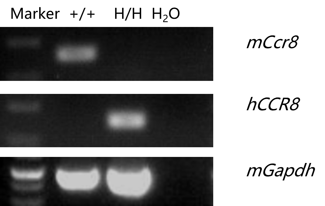
mRNA expression analysis
- Mouse Ccr8 mRNA was detectable in wild-type C57BL/6 mice.
- Human CCR8 mRNA was detectable only in homozygous B-hCCR8 mice but not in wild-type mice.
Strain specific analysis of CCR8 mRNA expression in wild-type C57BL/6 mice and homozygous B-hCCR8 mice by RT-PCR. Thymus RNA was isolated from wild-type C57BL/6 mice (+/+) and homozygous B-hCCR8 mice (H/H), then cDNA libraries were synthesized by reverse transcription, followed by PCR with mouse or human CCR8 primers.
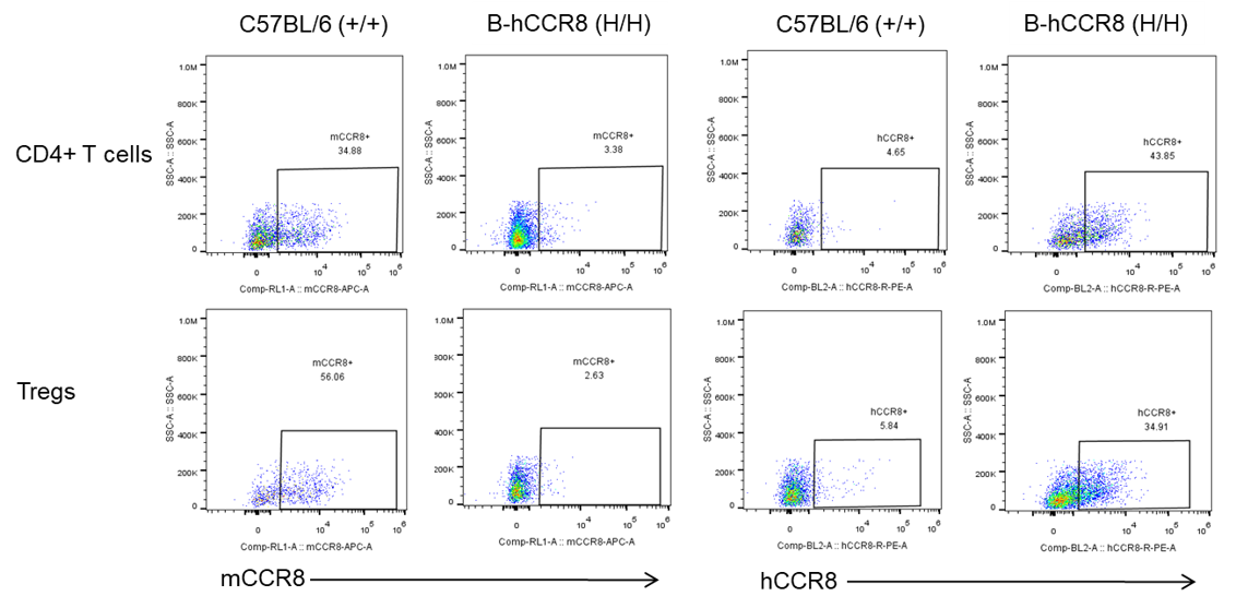
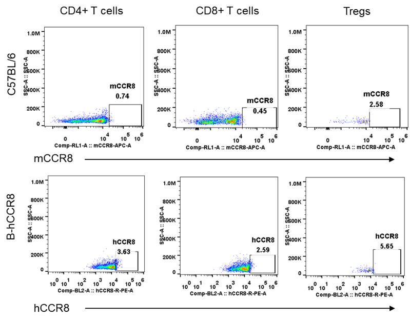
CCR8 Protein Expression in Tumor-infiltrating lymphocytes (TILs)
- Mouse Ccr8 was only detected on CD4+ T cells and Tregs in wild-type C57BL/6 mice.
- Human CCR8 was detected on CD4+ T cells and Tregs in B-hCCR8 mice, but not in wild-type C57BL/6 mice.
Strain specific CCR8 expression analysis in homozygous B-hCCR8 mice by FACS. MC38 cells were inoculated into wild-type C57BL/6 (+/+) and homozygous B-hCCR8 mice (H/H). Tumors were harvested at the endpoint of experiment, and the TILs were analyzed by flow cytometry.
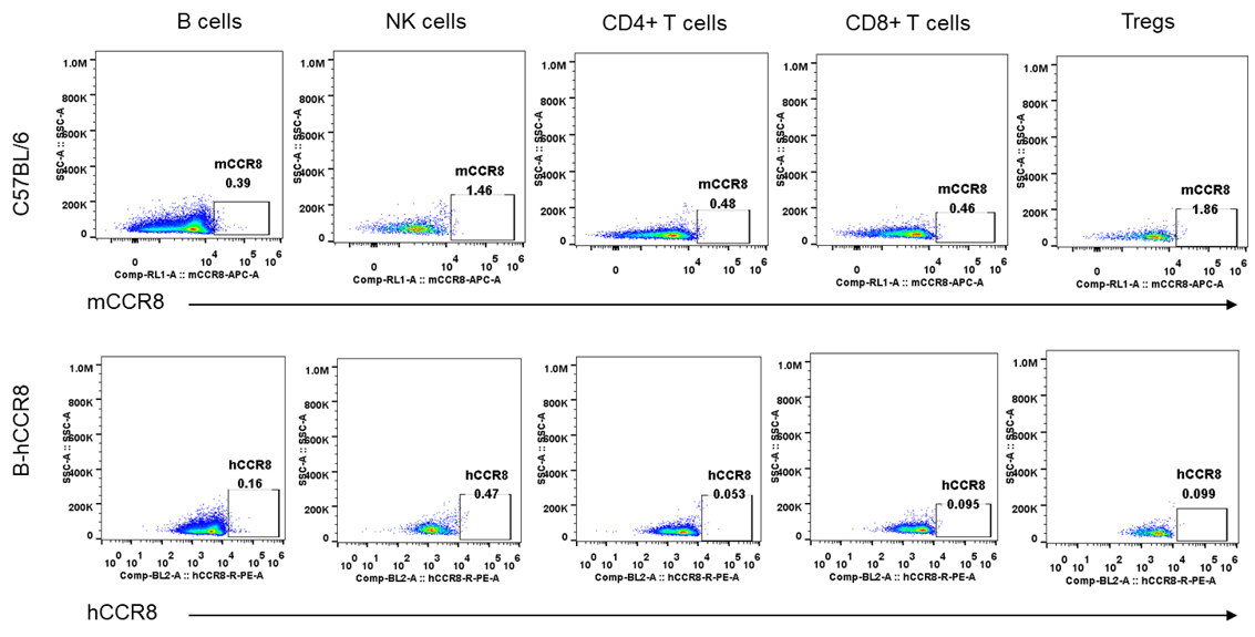
CCR8 Protein Expression in spleen
- Human and mouse CCR8 were not detected neither in splenocytes of wild-type mice nor B-hCCR8 mice separately.
Mouse and human CCR8 expression analysis in splenocytes. Splenocytes were collected from wild-type C57BL/6 mice and homozygous B-hCCR8 mice. CCR8 expression on B cells, NK cells, CD4+ T cells, CD8+ T cells and Tregs was analyzed by flow cytometry using species-specific anti-CCR8 antibodies.
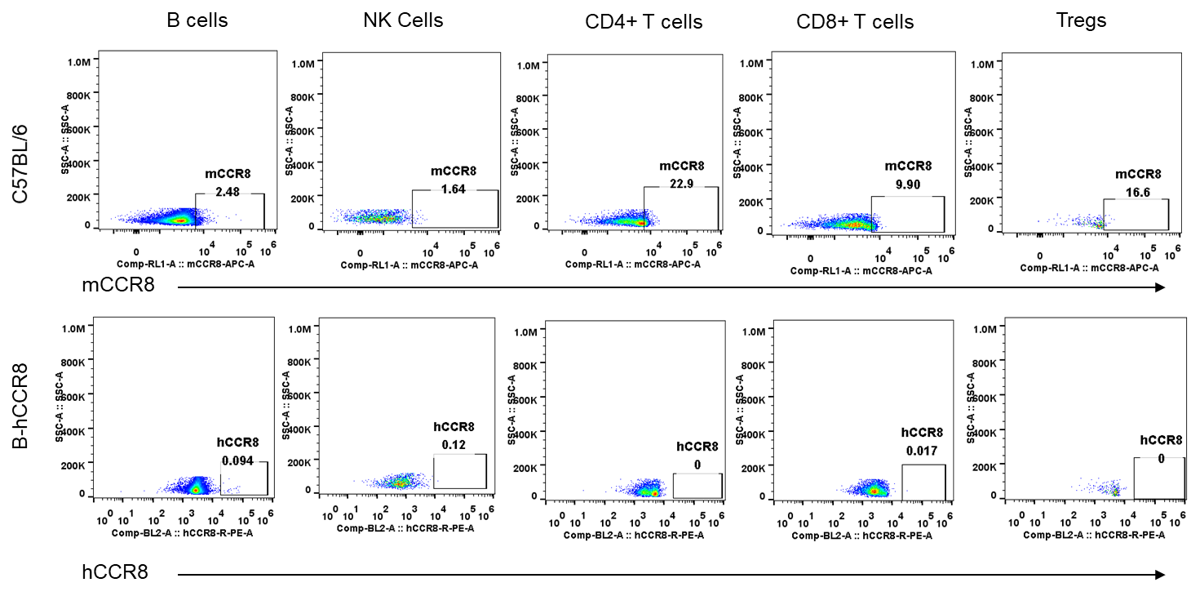
CCR8 Protein Expression in blood
- Human and mouse CCR8 were not detected neither in blood cells of wild-type mice nor B-hCCR8 mice separately.
Mouse and human CCR8 expression analysis in blood. Blood were collected from wild-type C57BL/6 mice and homozygous B-hCCR8 mice. CCR8 expression on B cells, NK cells, CD4+ T cells, CD8+ T cells and Tregs was analyzed by flow cytometry using species-specific anti-CCR8 antibodies.
CCR8 Protein Expression in thymus
- Human and mouse CCR8 were not expressed neither in thymus cells of wild-type mice nor B-hCCR8 mice separately.
Mouse and human CCR8 expression analysis in thymus. Thymus were collected from wild-type C57BL/6 mice and homozygous B-hCCR8 mice. CCR8 expression on CD4+ T cells, CD8+ T cells and Tregs was analyzed by flow cytometry using species-specific anti-CCR8 antibodies.
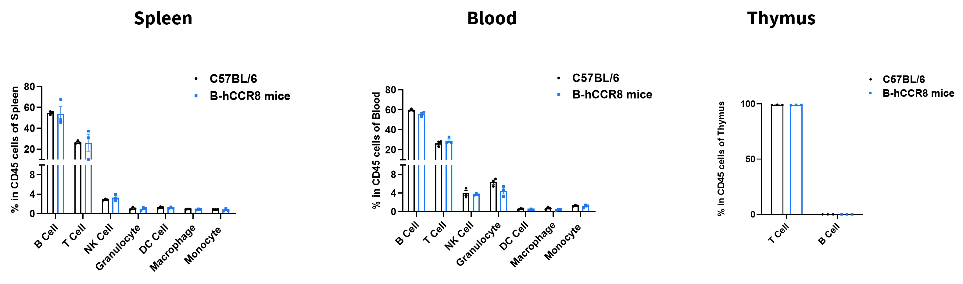
Analysis of Leukocyte Subpopulations
- The percentages of T cells, B cells, NK cells, DCs, granulocytes, monocytes, and macrophages in homozygous B-hCCR8 mice are similar to those in C57BL/6JNifdc mice.
- Humanization of CCR8 does not affect normal immune cell development or splenic distribution.
Analysis of leukocyte subpopulations by flow cytometry in immune organs and blood. Splenocytes, peripheral blood, and thymus were isolated from female C57BL/6JNifdc and B-hCCR8 mice (female, 8-week-old, n = 3). Single live cells were gated on the CD45⁺ population and analyzed by flow cytometry as indicated. Values are expressed as mean ± SEM.
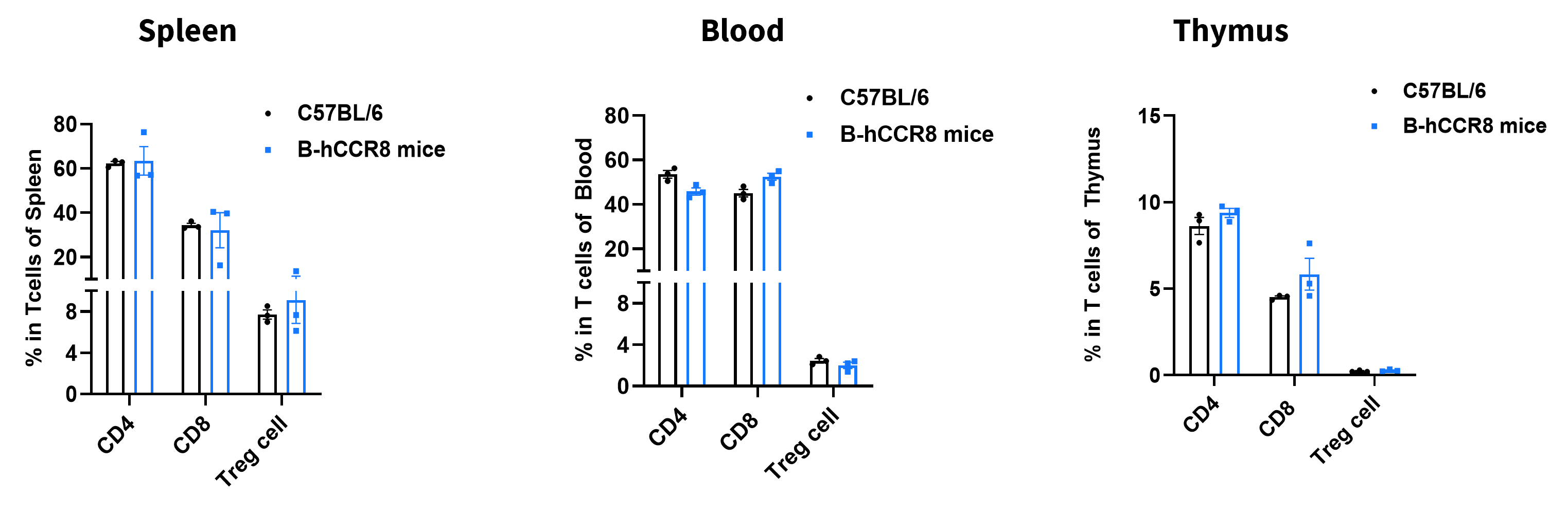
Analysis of T Cell Subpopulations
- The proportions of CD4⁺ T cells, CD8⁺ T cells, and Tregs in homozygous B-hCCR8 mice are comparable to those in C57BL/6JNifdc mice.
- Humanization of CCR8 does not affect normal T cell development, differentiation, or splenic distribution.
Analysis of T-cell subpopulations by flow cytometry in immune organs and blood. Splenocytes, peripheral blood, and thymus were isolated from female C57BL/6JNifdc and B-hCCR8 mice (female, 8-week-old, n = 3). Single live cells were gated on the CD3⁺ T-cell population and analyzed by flow cytometry as indicated. Values are expressed as mean ± SEM.
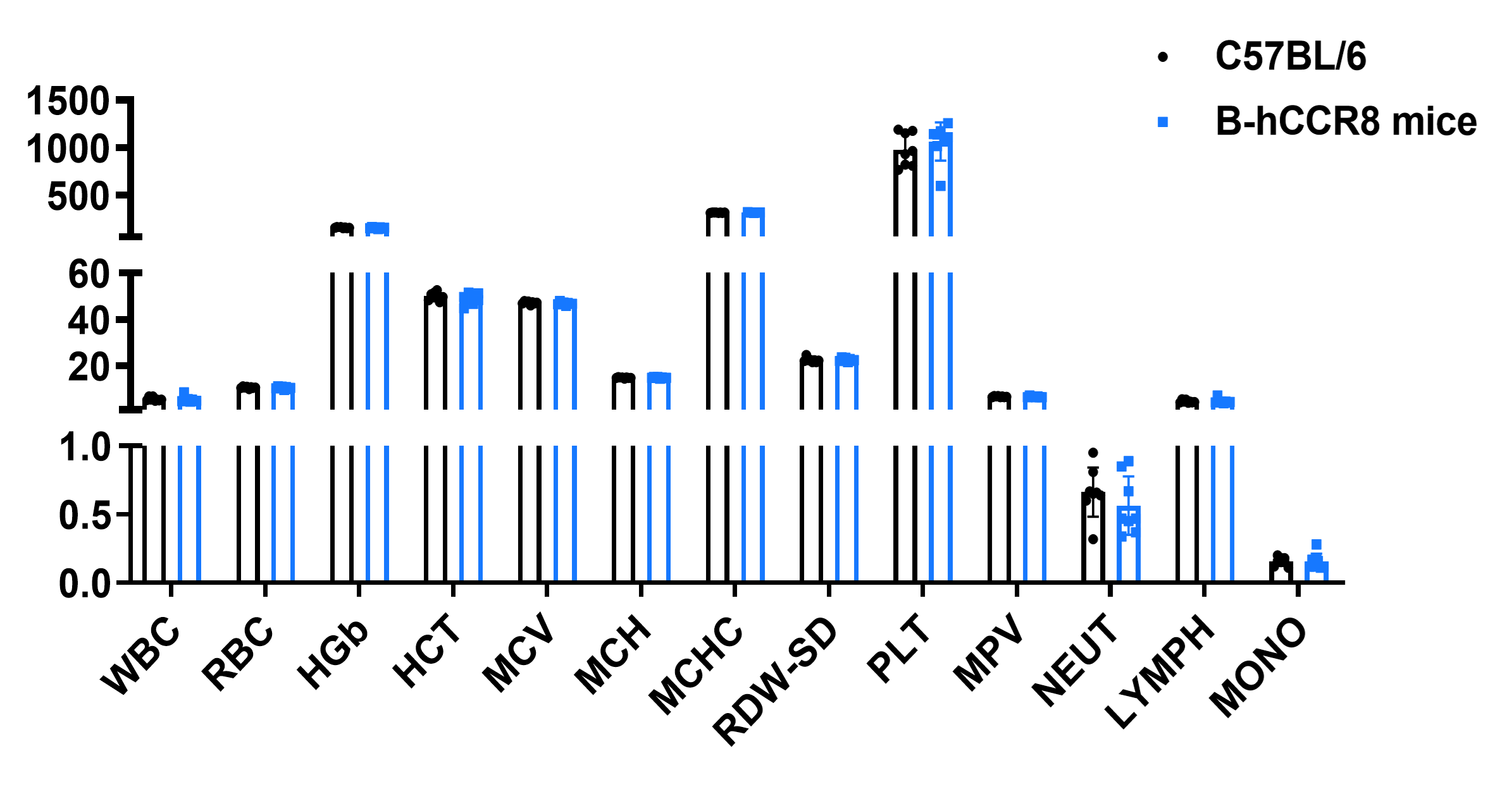
Hematology Analysis
- No significant differences were observed compared with wild-type mice.
Complete blood count (CBC) of B-hCCR8 mice. Values are expressed as mean ± SD.
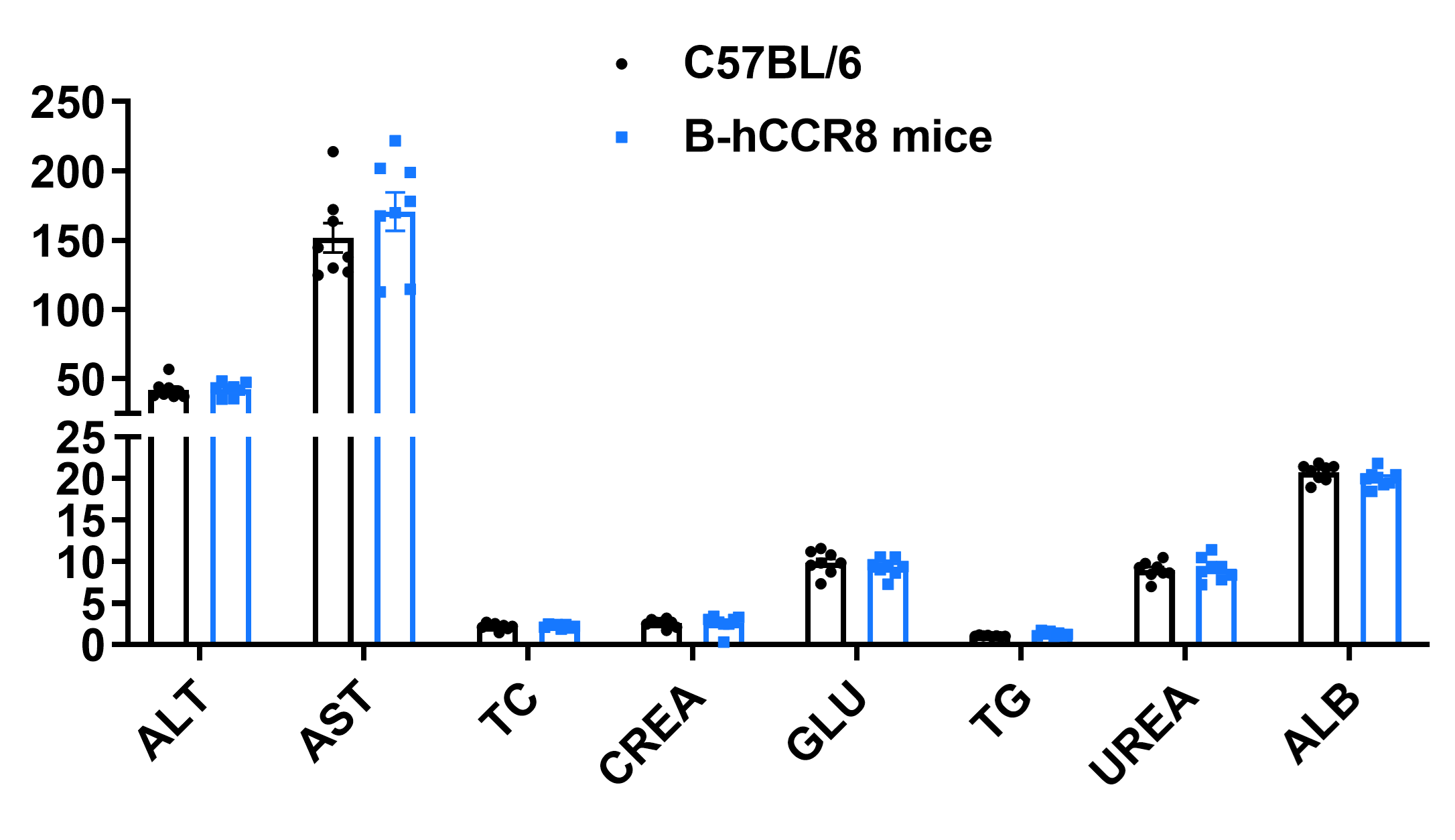
Blood Biochemical Analysis
- No significant differences were observed compared with wild-type mice.
Blood biochemical parameters of B-hCCR8 mice are shown. Values are expressed as mean ± SD.
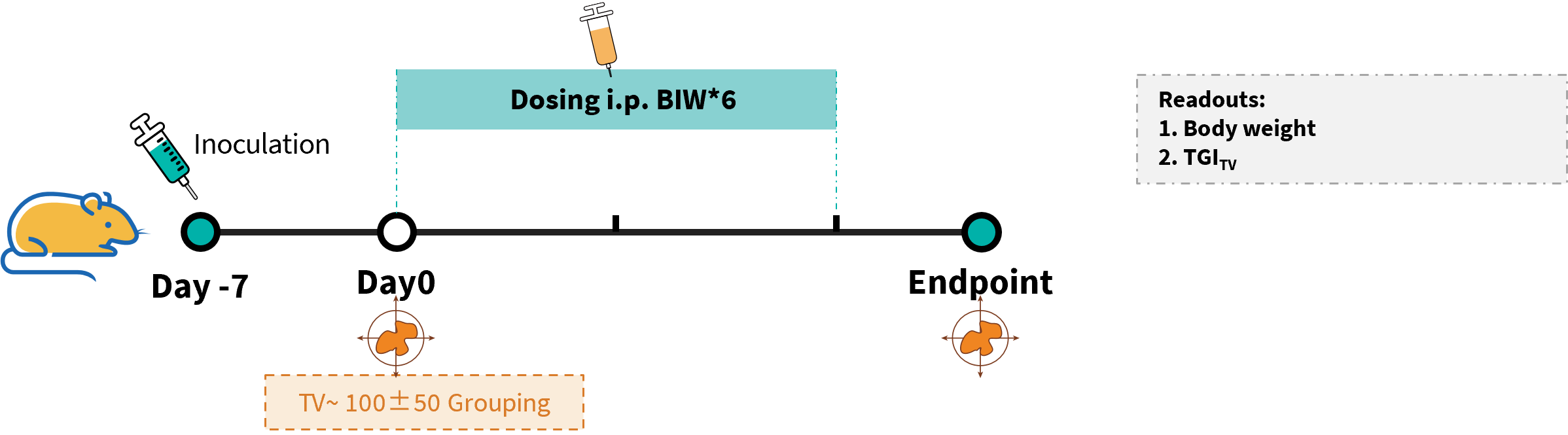
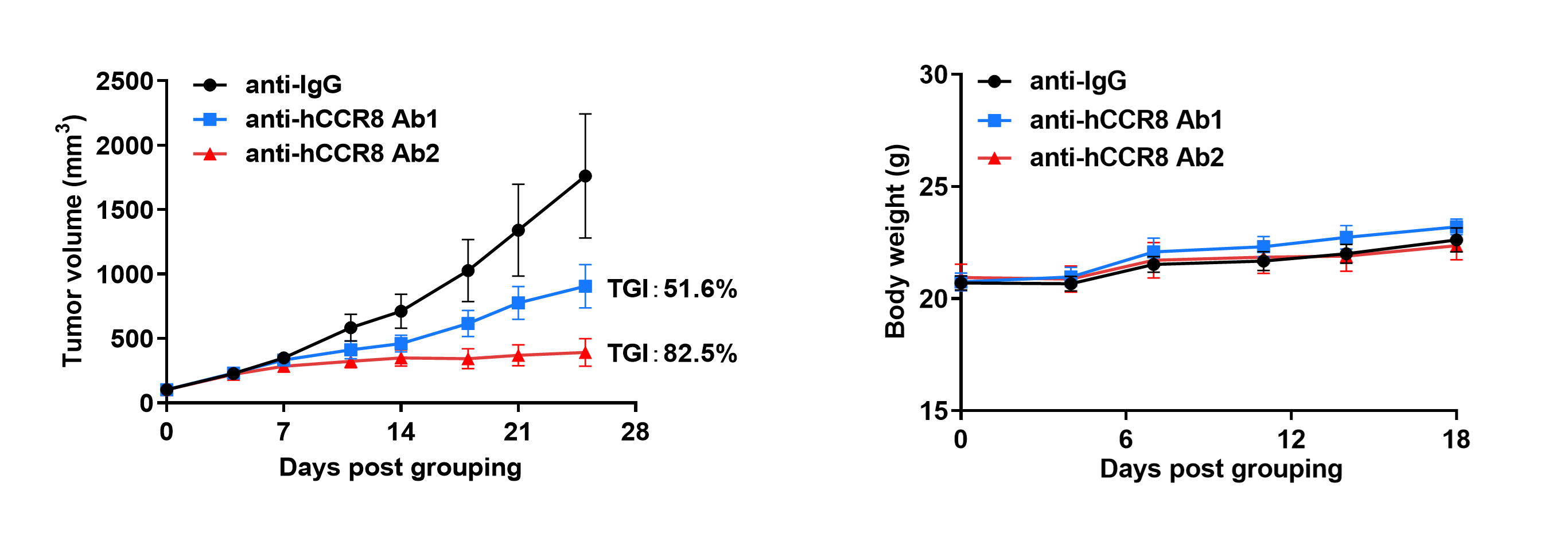
Efficacy evalsuation of anti-CCR8 antibody in the Treatment of the Subcutaneous MC38 Model in B-hCCR8 mice in vivo
Establishment of a MC38 model and in vivo efficacy study of anti-CCR8 antibody. Murine colon cancer MC38 cells were subcutaneously implanted into homozygous B-hCCR8 mice (female, 7–week-old, n=6). Mice were grouped when tumor volume reached approximately 100 mm³, at which time they were injected intraperitoneally with anti-human CCR8 antibodies (in house).
- Anti-human CCR8 antibodies were efficacious in controlling tumor growth in B-hCCR8 mice.
- B-hCCR8 mice provide a powerful preclinical model for in vivo evalsuation of anti-human CCR8 antibodies.
Efficacy of anti-human CCR8 antibodies in B-hCCR8 mice. (A) Tumor growth curves. (B) Body weight changes during treatment. Values are expressed as mean ± SEM.
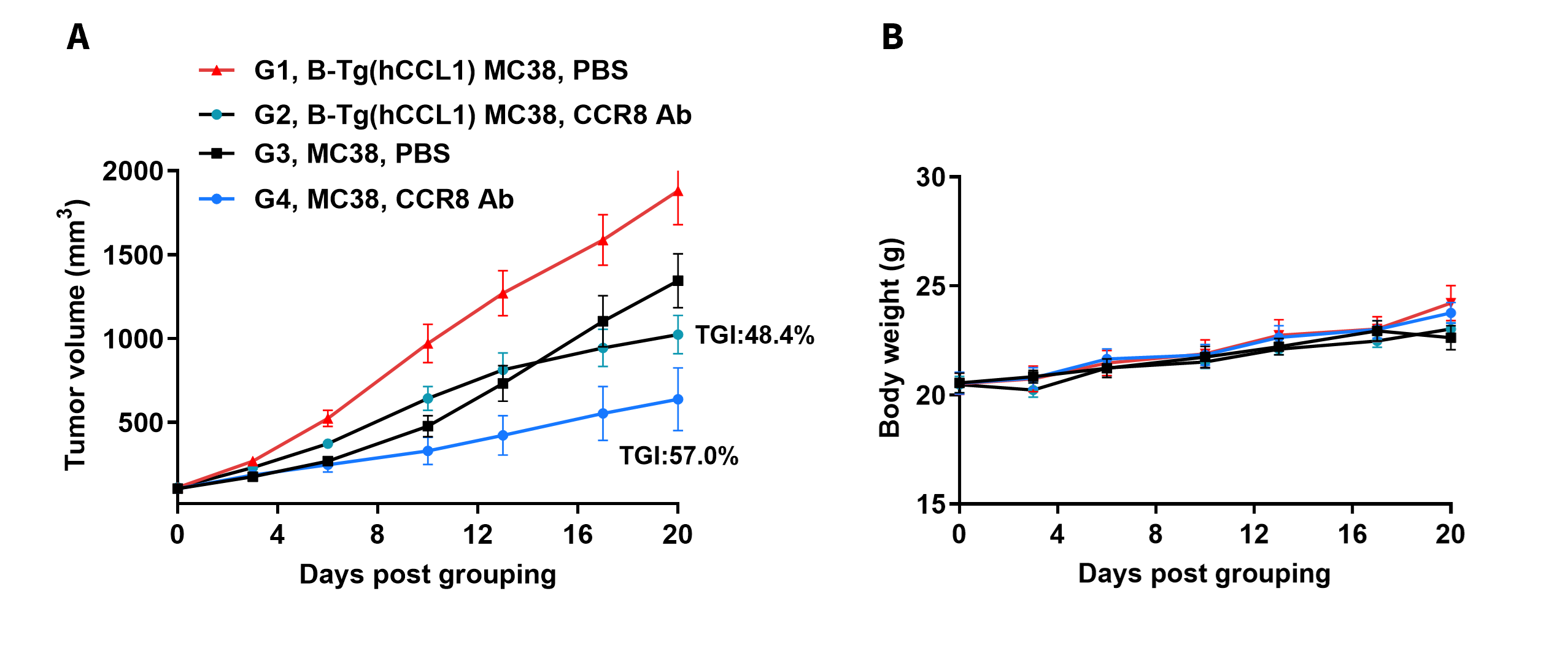
Establishment of a MC38 model and in vivo efficacy study of anti-CCR8 antibody. Wild-type MC38 cells and B-Tg(hCCL1) MC38 cells (Clone: 2-C12) were subcutaneously implanted into homozygous B-hCCR8 mice (female, 8–week-old, n=7). Mice were grouped when tumor volume reached approximately 100 mm³, at which time they were injected intraperitoneally with anti-human CCR8 antibodies from cooperation company.
Efficacy evalsuation of anti-CCR8 antibody in the Treatment of the Subcutaneous MC38 Model in B-hCCR8 mice in vivo
- Anti-human CCR8 antibodies were efficacious in controlling tumor growth in B-hCCR8 mice.
- B-hCCR8 mice provide a powerful preclinical model for in vivo evalsuation of anti-human CCR8 antibodies.
Efficacy of anti-human CCR8 antibodies in B-hCCR8 mice. (A) Tumor growth curves. (B) Body weight changes during treatment. Values are expressed as mean ± SEM.
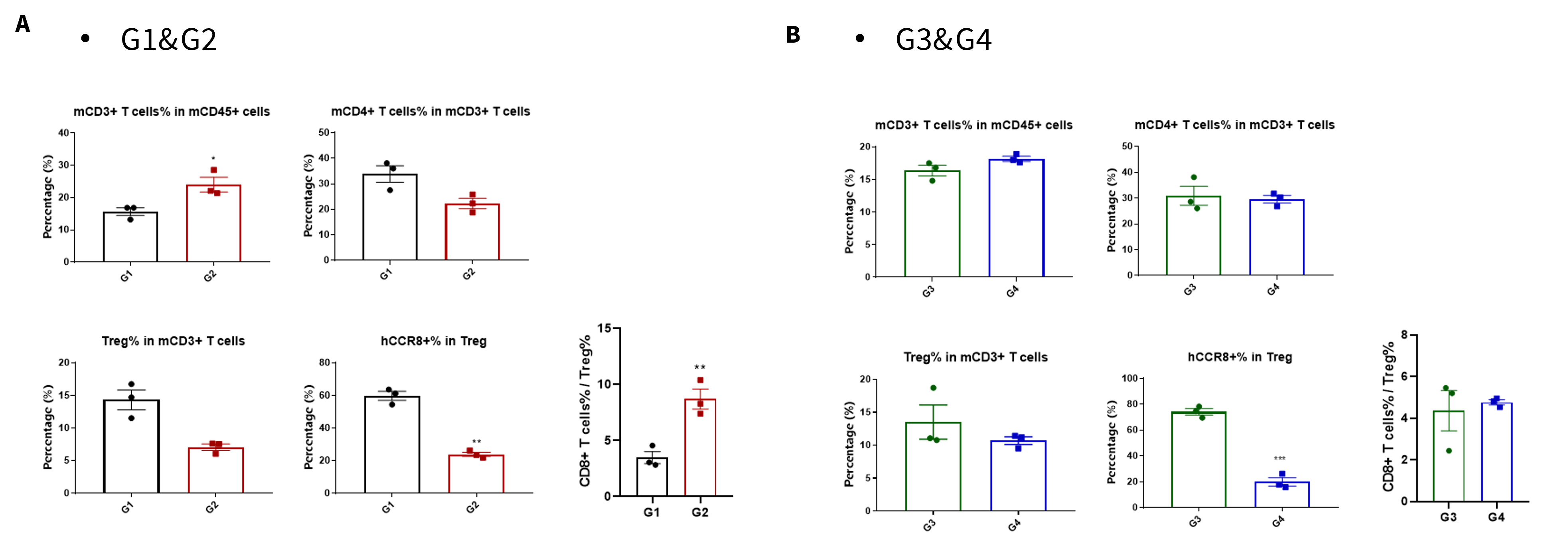
Analysis of tumor infiltrates lymphocytes by FACS. TILs were harvested at the endpoint of the experiment and flow cytometry analysis was performed to assess the leukocyte subpopulations. The percent of CCR8+ Treg cells were significant decrease in anti-human CCR8 antibody treatment group (G2, G4), and the ratio CD8+ T cells to Treg cells increased significantly in G2, but not in G4. (*p<0.05, **p<0.01, ***p<0.001, ****p<0.0001)
* When publishing results obtained using this animal model, please acknowledge the source as follows: The animal model [B-hCCR8 mice] (Cat# 110096) was purchased from Biocytogen.